2026 Top Medical Imaging Equipment Types for Global Buyers

Global buyers entering the 2026 market need more than a catalogue of impressive machines. They need a practical view of clinical demand, operating costs, service access, and long-term upgrade potential. This guide examines the leading medical imaging equipment types, including digital X-ray, ultrasound, CT, MRI, mammography, and PET/CT systems. Each modality serves different patients, workflows, budgets, and facility sizes.

Real-world purchasing rarely follows a perfect checklist. A compact ultrasound system may outperform a larger unit in rural clinics. A high-field MRI can deliver exceptional detail, yet demand stronger infrastructure and trained staff. CT scanners offer speed, but buyers must examine dose-management tools, cooling requirements, and maintenance response times. Small details matter. So does local experience.

Dr. Curt Langlotz, a Stanford radiologist and artificial intelligence researcher, stated, “AI will not replace radiologists, but radiologists who use AI will replace those who don’t.” His observation reflects a wider shift toward connected imaging platforms, automated workflows, and decision-support software. However, technology alone does not guarantee better care. Buyers should verify image quality, cybersecurity controls, interoperability, warranty terms, and application training before signing contracts. Independent evaluations and transparent vendor documentation remain essential.

This overview is designed for distributors, hospitals, diagnostic centers, and public-health planners comparing global options. It considers clinical versatility, total cost of ownership, installation demands, and supplier reliability. Some recommendations may change as software, regulations, and procurement conditions evolve. That uncertainty deserves attention. A careful buyer should question every attractive specification.

2026 Top Medical Imaging Equipment Types for Global Buyers

Medical Imaging Equipment: Definition, Scope, and Core Functions

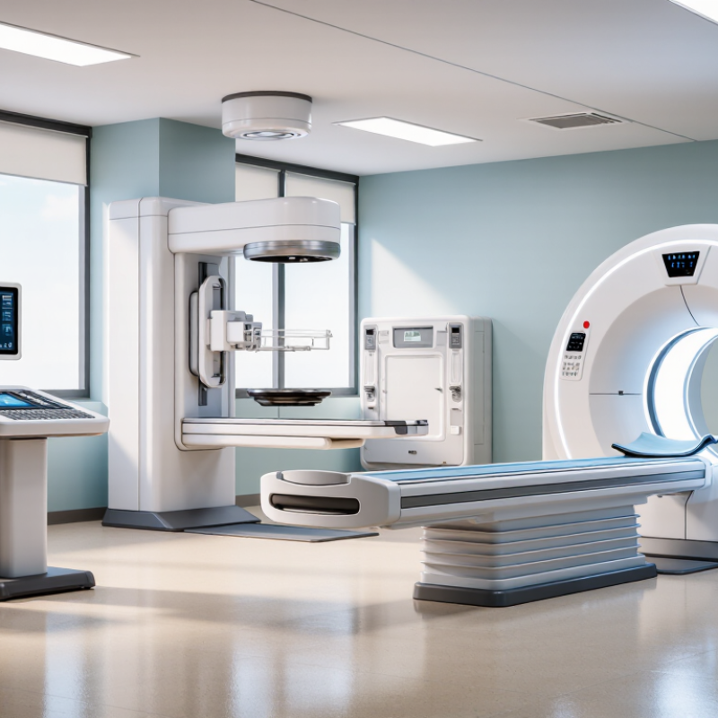
Medical imaging equipment refers to systems that create visual information about the human body. It supports screening, diagnosis, treatment planning, and follow-up care. Its scope includes X-ray, computed tomography, magnetic resonance imaging, ultrasound, and nuclear imaging systems. Each modality uses different physical principles and produces different clinical information.

Core functions include image acquisition, processing, storage, transmission, and review. X-ray systems capture dense structures quickly. CT combines multiple views into cross-sectional images. MRI offers strong soft-tissue contrast without ionizing radiation. Ultrasound provides real-time imaging at the bedside. Nuclear imaging can show biological activity, not only anatomy.

These differences affect installation, staffing, safety controls, and maintenance.

For global buyers, evaluation must extend beyond image quality. Check radiation protection, electrical safety, calibration procedures, data interoperability, and regulatory documentation. A rural clinic may prioritize low power demand and simple maintenance. A major hospital may need high patient throughput and integration with existing systems. Service response, operator training, spare parts, and language support also matter. A scanner can look impressive on paper and still frustrate staff. Real-world workflow often exposes overlooked weaknesses. Procurement teams should test representative cases, review quality assurance records, and involve clinicians and technicians. No device fits every setting. Careful selection remains partly technical and partly human.

Major Equipment Types: X-Ray, CT, MRI, Ultrasound, and Nuclear Imaging

2026 Top Medical Imaging Equipment Types for Global Buyers

X-ray remains the practical entry point for many hospitals. It supports chest, bone, dental, and emergency imaging with relatively modest space requirements. The World Health Organization’s Global Atlas of Medical Devices highlights major gaps in equipment access and maintenance across lower-resource settings. Buyers should inspect detector durability, service response, radiation controls, and replacement-part availability. Low purchase cost can become expensive downtime.

CT provides rapid cross-sectional imaging for trauma, stroke, and cancer assessment. However, it requires higher shielding, electrical capacity, cooling, and dose-management discipline. OECD Health Statistics 2024 reports that equipment density varies sharply between countries; Japan has more than 100 CT scanners per million people. MRI avoids ionizing radiation and offers strong soft-tissue contrast. It also demands careful siting, stable power, trained operators, and longer examination times. No modality wins everywhere.

Ultrasound is compact and adaptable, especially for obstetrics, abdominal studies, and bedside care. Its image quality depends heavily on operator skill, probe selection, and clinical workflow. Nuclear imaging adds physiological information through radiotracers, supporting oncology, cardiology, and thyroid evaluation. The IAEA reports that nuclear medicine services perform roughly 40 million procedures globally each year, but access remains uneven. Buyers must assess isotope supply, licensing, waste handling, and camera sensitivity. A spreadsheet cannot capture every local constraint. My caution is simple: specifications may look complete, while training and service planning remain unfinished.

2026 Top Medical Imaging Equipment Types for Global Buyers

Typical patient-facing examination time by major medical imaging modality

Representative clinical workflow benchmarks indicate that X-Ray examinations are usually the fastest, while nuclear imaging and MRI generally require more patient time. Actual duration varies according to examination type, body region, contrast use, patient condition, and facility workflow. These figures describe typical examination time and are not market-share estimates.

Key Specifications for Comparing Imaging Systems Across Markets

2026 Top Medical Imaging Equipment Types for Global Buyers

Comparing imaging systems requires more than counting features. CT buyers should review slice configuration, rotation speed, dose-control tools, detector coverage, and reconstruction quality. MRI evaluations should include field strength, gradient performance, coil design, scan time, and cooling requirements. For ultrasound, probe frequency, frame rate, penetration, image uniformity, and battery life matter in daily use. A higher specification is not always better.

Market conditions can change the right choice. A powerful CT system may struggle where electrical capacity is unstable. An MRI unit may require temperature control, shielding, trained engineers, and dependable spare parts. Check DICOM compatibility, cybersecurity controls, warranty coverage, installation access, and service response times. Total cost includes electricity, consumables, software updates, and staff training. Procurement teams should request independent image-quality tests, not rely only on supplier demonstrations. Small oversights become expensive.

Tips: Compare systems with the same clinical protocol and patient workload. Ask for measured dose, uptime records, and sample images from similar facilities. Review local regulatory requirements before purchase. Leave space for maintenance access. Be careful with projected patient volumes; early estimates are often optimistic. A practical system with reliable support can outperform a technically superior unit that remains idle. Test the workflow with real operators, because specifications rarely reveal every awkward step.

2026 Top Medical Imaging Equipment Types for Global Buyers - Key Specifications for Comparing Imaging Systems Across Markets
The specifications below are representative procurement ranges for commonly configured clinical systems. Actual performance depends on system configuration, local regulations, installed options, examination protocols, and site conditions.
Equipment Type Primary Clinical Applications Key Technical Specifications to Compare Typical Configuration Ranges Patient and Workflow Considerations Infrastructure Requirements Recommended Buyer Evaluation Points
Digital Radiography (DR) General radiography, emergency imaging, chest examinations, skeletal studies, mobile ward imaging, and trauma assessment. Detector technology, detector size, pixel pitch, spatial resolution, detective quantum efficiency (DQE), tube voltage range, tube current, exposure automation, image stitching, and wireless detector options. 43 × 43 cm detector 17 × 17 in detector Approximately 40–150 kVp Pixel pitch commonly 100–200 µm Fixed or portable configurations Fast examinations and high patient throughput. Detector durability, battery replacement, cleaning procedures, weight, and drop resistance are important for mobile use. Shielded radiography room for fixed systems, stable electrical supply, radiation protection, network connectivity, and adequate space for patient positioning. Portable systems require charging and storage areas. Compare image quality at clinically relevant dose levels, detector warranty terms, interoperability with RIS/PACS, exposure consistency, software workflow, service response, and availability of replacement detectors.
Computed Tomography (CT) Trauma, stroke, oncology, chest and abdominal imaging, angiography, cardiac studies, virtual colonography, and image-guided procedures. Detector row and slice configuration, z-axis coverage, rotation time, spatial and low-contrast resolution, dose modulation, iterative or deep-learning reconstruction, tube heat capacity, spectral or dual-energy capability, and table load. 16–128 detector rows Approximately 0.5–0.625 mm detector collimation About 0.28–0.5 s rotation time Approximately 70–140 kVp Typical gantry aperture near 70 cm High throughput and broad clinical versatility. Radiation dose management, patient centering, emergency access, bariatric capacity, and contrast-injection workflow require close evaluation. Dedicated shielded room, high-capacity electrical supply, heat removal or air conditioning, structural assessment, contrast injector provisions, emergency equipment, and reliable data-network infrastructure. Evaluate dose-length-product performance, pediatric protocols, scan speed, tube cooling, uptime, reconstruction speed, cardiac capability, spectral imaging value, service coverage, and total cost of ownership.
Magnetic Resonance Imaging (MRI) Neurology, spine, musculoskeletal imaging, abdominal and pelvic studies, angiography, oncology, cardiac imaging, and functional or diffusion imaging. Field strength, gradient amplitude and slew rate, maximum field of view, bore diameter, RF channels, parallel imaging, acceleration methods, acoustic noise, scan speed, SAR management, and coil selection. 1.5 T or 3.0 T clinical systems Approximately 60–70 cm bore Common gradient amplitude 30–45 mT/m Multiple dedicated receive coils Typical examination time 15–60 minutes No ionizing radiation, but screening for implants and metal is mandatory. Patient comfort, claustrophobia management, motion control, acoustic noise, and anesthesia compatibility affect workflow. RF-shielded room, quench-pipe planning where applicable, magnetic-zone controls, vibration management, stable power, cooling, non-ferromagnetic accessories, and controlled access. Compare clinical protocols, artifact reduction, implant safety documentation, gradient performance, coil ergonomics, helium or cooling requirements, uptime, software licensing, and local technical support.
Diagnostic Ultrasound Abdominal, obstetric, gynecologic, vascular, cardiac, breast, thyroid, musculoskeletal, emergency, and point-of-care examinations. Probe portfolio, frequency bandwidth, frame rate, penetration, Doppler sensitivity, color and power Doppler performance, elastography, 3D/4D capability, contrast-enhanced ultrasound support, monitor quality, and AI-assisted workflow. Approximately 1–18 MHz probe range Convex, linear, phased-array, endocavitary probes Cart-based or handheld systems 2D, Doppler, and optional 3D/4D imaging No ionizing radiation and generally portable. Image quality is highly operator-dependent. Probe cleaning, infection control, ergonomics, battery life, and data export are key operational issues. Standard electrical supply, examination space, secure network connection, probe disinfection facilities, and optional battery-charging infrastructure for portable units. Compare clinical presets, Doppler sensitivity, probe durability, image uniformity, cybersecurity, DICOM compatibility, software upgrade policy, replacement-probe cost, and user training.
Mammography Breast cancer screening, diagnostic mammography, magnification imaging, implant evaluation, and localization or biopsy guidance when configured for intervention. Detector type, detector size, pixel pitch, image quality, automatic exposure control, compression system, tomosynthesis performance, dose indicators, biopsy accuracy, and positioning ergonomics. Typical detector area near 24 × 30 cm Approximately 20–40 kV tube voltage Pixel pitch commonly about 70–100 µm 2D and optional digital breast tomosynthesis Requires careful positioning and controlled compression. Patient communication, examination speed, repeat-rate monitoring, infection control, and accessibility are important for screening programs. Shielded room, stable power, dedicated positioning space, radiation protection, image-review workstation, and sufficient network capacity for large tomosynthesis datasets. Compare dose and image quality, tomosynthesis acquisition time, reconstruction speed, compression control, artifact performance, biopsy workflow, regulatory approvals, and quality-control tools.
Nuclear Medicine SPECT/CT Bone, cardiac perfusion, endocrine, renal, infection, inflammation, and selected oncology examinations using radiotracers. Detector crystal type, energy resolution, sensitivity, spatial resolution, number of detector heads, CT capability, attenuation correction, motion correction, quantitative software, and collimator options. Usually dual-head gamma-camera geometry Low- and medium-energy collimator options Integrated low-dose CT commonly available Energy resolution often evaluated near 140 keV Examination time can be longer than conventional radiography or CT. Radiotracer handling, patient scheduling, contamination control, and uptake-time management influence department efficiency. Radiation-controlled area, radiopharmacy or tracer-handling facilities, shielding, contamination-monitoring equipment, secure isotope storage, and appropriate waste-management procedures. Evaluate count sensitivity, attenuation-correction accuracy, quantitative capability, CT dose, detector uniformity, software compatibility, isotope logistics, service support, and regulatory compliance.
PET/CT Oncology staging and treatment response, selected neurologic studies, cardiac viability, and whole-body molecular imaging. Detector material, time-of-flight performance, sensitivity, spatial resolution, axial field of view, coincidence timing, CT detector configuration, attenuation correction, motion correction, and quantitative standardization. Typical PET axial coverage about 15–30 cm Time-of-flight systems commonly use sub-nanosecond timing Integrated CT commonly 16–128 detector rows Typical whole-body acquisition about 15–30 minutes Requires radiotracer preparation and strict patient scheduling. Occupancy time, uptake-room capacity, patient motion, glucose management, and radiation safety affect throughput. Shielded uptake and scanning areas, radiotracer supply chain, dose calibrator, contamination controls, radiation monitoring, cooling, high-capacity power, and specialized service support. Compare sensitivity, quantitative accuracy, timing performance, scan range, CT dose, motion correction, radiotracer compatibility, uptime, regulatory requirements, and isotope availability in the target market.
Fluoroscopy and Digital Angiography Interventional radiology, vascular procedures, cardiac procedures, gastrointestinal studies, pain management, orthopedic surgery, and image-guided treatment. Detector size, pulse rate, frame rate, flat-panel performance, field of view, C-arm geometry, table movement, magnification, roadmapping, dose monitoring, image-processing tools, and maximum patient weight. Flat-panel detectors commonly 20–30 cm or 30–40 cm class Pulsed fluoroscopy commonly 7.5–30 frames/s Single-plane or biplane configurations Fixed-room or mobile C-arm systems Supports real-time guidance but may involve extended radiation exposure. Table access, sterile workflow, staff protection, procedure-room turnover, and emergency access are critical. Shielded procedure room, ceiling or table-mounted protective barriers, high-capacity power, heat management, anesthesia and monitoring interfaces, sterile-area planning, and network connectivity. Compare dose-rate control, image quality at low dose, detector coverage, C-arm flexibility, table load, collision avoidance, 3D imaging, procedure software, service response, and staff training.
Dental Cone-Beam CT (CBCT) Dental implant planning, oral and maxillofacial surgery, orthodontics, endodontics, temporomandibular-joint assessment, and selected ear-nose-throat applications. Field of view, voxel size, tube voltage and current, artifact reduction, reconstruction software, dose optimization, patient positioning, cephalometric capability, and image-export formats. Field of view commonly about 4–20 cm Voxel sizes often approximately 0.08–0.4 mm Approximately 60–100 kVp Seated, standing, or supine designs Compact workflow and high-resolution dental imaging. Patient motion, metal artifacts, pediatric protocols, positioning repeat rates, and accessibility should be assessed. Room shielding based on local regulations, stable power, adequate operator visibility, network connectivity, and sufficient space for patient positioning and infection-control procedures. Compare dose by field of view, artifact reduction, voxel-size usefulness, software interoperability, implant-planning tools, regulatory status, maintenance requirements, and training resources.
Note: Buyers should verify country-specific electrical standards, radiation regulations, medical-device registration, cybersecurity requirements, warranty conditions, preventive-maintenance schedules, and availability of qualified service personnel before purchase.

Clinical Applications and Departmental Equipment Requirements

2026 Top Medical Imaging Equipment Types for Global Buyers

Clinical applications should guide every imaging purchase, not catalogue popularity. Radiography remains essential for emergency rooms, orthopedic clinics, and mobile services. CT supports trauma, stroke, oncology, and cardiac assessment. MRI serves neurology, musculoskeletal care, and soft-tissue diagnosis, but requires stronger infrastructure and longer examination times. Ultrasound fits obstetrics, abdominal care, intensive care, and primary clinics. Nuclear imaging and radiotherapy equipment demand specialized shielding, licensing, and highly trained teams.

The OECD Health Statistics 2024 report shows major differences in MRI availability. Japan reported more than 50 MRI units per million people, while several countries recorded fewer than 10. This gap reflects budgets, workforce capacity, and referral systems, not only demand. IAEA guidance also stresses quality assurance, radiation protection, and maintenance planning. A department may purchase advanced equipment yet underuse it. That is a costly and realistic weakness.

Tips: Map equipment to patient volume, referral patterns, room size, and local service access. Check power stability, cooling, shielding, software compatibility, and spare-part support before ordering. Ask for acceptance testing and staff training. Do not treat artificial intelligence as a replacement for clinical judgment. Procurement teams should review OECD data, IAEA technical guidance, and national regulations together. Estimates can age quickly. Recheck them before final approval.

Global Buyer Considerations: Compliance, Cost, Support, and Integration

Global buyers assessing medical imaging equipment in 2026 should examine compliance before comparing prices. Each market may require different registrations, safety testing, labeling, and data protection controls. Request complete technical files, electrical safety reports, clinical documentation, and software security records. Paperwork matters. A missing certificate can delay installation for weeks.

Purchase cost is only one part of financial planning. Buyers should calculate installation, room adjustments, operator training, maintenance, consumables, and eventual upgrades. A cheaper system may demand expensive infrastructure changes. I have seen projects underestimate cooling, power stability, and shielding requirements. Those oversights can disrupt budgets and patient services. Cost models should include local service rates and expected downtime.

Reliable support needs measurable commitments. Ask about response times, spare-part availability, remote diagnostics, and engineer coverage near the facility. Support matters. Integration also deserves practical testing before purchase. Confirm compatibility with existing imaging archives, hospital information systems, reporting tools, and cybersecurity policies. Use realistic test studies, not only vendor demonstrations. Integration fails quietly when worklists, patient identifiers, or image formats behave differently in daily use. Buyers should also verify staff training plans and maintenance instructions. No evaluation is perfect, and technical teams may overlook workflow friction during a short demonstration. A supervised pilot can reveal these weaknesses before a long-term contract.

Go to Top
Have Questions? Call (888) 602-pros